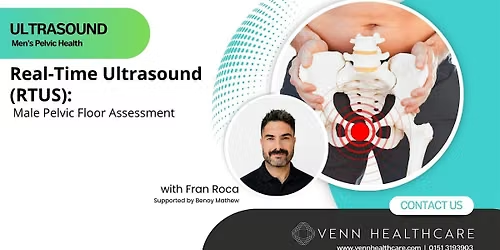
Real-Time Ultrasound (RTUS) in Male Pelvic Floor Assessment

About this Event
Who is this course for?
This workshop is designed for healthcare professionals involved in the assessment and rehabilitation of men with pelvic floor dysfunction and lower urinary tract symptoms. It is suitable for:
- Physiotherapists working in pelvic health
- Urologist looking to intergrate ultrasound into their clinical workplan
- Specialist pelvic health physiotherapists wishing to incorporate RTUS into clinical practice
- Continence nurses and urology nurse specialists
- Advanced clinical practitioners working in men's health
- Healthcare professionals involved in the rehabilitation of men following prostate cancer treatment or prostate surgery
- Clinicians managing patients with chronic pelvic pain syndrome (CPPS), lower urinary tract symptoms (LUTS), urinary incontinence, or pelvic floor dysfunction
What will the course offer?
This practical workshop introduces clinicians to the use of transabdominal real-time ultrasound (RTUS) as a non-invasive assessment and biofeedback tool for pelvic floor rehabilitation in men. Combining evidence-informed theory with live demonstration and hands-on practice, participants will develop the skills to acquire and interpret ultrasound images, identify normal and dysfunctional pelvic floor movement patterns, measure bladder volume and post-void residual, and apply RTUS to support patient education and rehabilitation.
Clinical applications will focus on the management of post-prostatectomy urinary incontinence, chronic pelvic pain syndrome (CPPS), and lower urinary tract symptoms (LUTS), with an emphasis on integrating RTUS into everyday clinical practice.
Learning Objectives
By the end of this workshop, participants will be able to:
- Explain the basic principles of diagnostic ultrasound, including safe operation, image orientation, and infection prevention procedures.
- Identify key pelvic structures on suprapubic transabdominal RTUS, including the bladder, bladder neck, urethra, prostate, and pelvic floor muscles.
- Recognise normal and dysfunctional pelvic floor movement patterns, including lift, bulge, overactivity, underactivity, poor coordination, and breath-related movement.
- Measure bladder volume and post-void residual using RTUS and interpret findings within a clinical context.
- Use RTUS as a real-time biofeedback tool to facilitate pelvic floor muscle training and optimise patient engagement.
- Apply RTUS findings to the assessment and management of men with post-prostatectomy urinary incontinence, chronic pelvic pain syndrome (CPPS), and lower urinary tract symptoms (LUTS).
- Demonstrate appropriate probe placement, image acquisition, and effective patient cueing during practical scanning.
Agenda
18:30–18:40 - Welcome & Outcomes
• Identify structures on suprapubic RTUS
• Recognise patterns
• Measure bladder volume
• Apply RTUS as biofeedback
• Link findings to post-prostatectomy, CPPS, LUTS
18:40–18.55 - US Basics
• What is ultrasound?
• The machine and reading the image
• Safety and hygiene: gel, probe covers, cleaning protocols
18:55–19:15 - Theory 1 Anatomy on RTUS
• Bladder, bladder neck, urethra, prostate and PFM.
19:15–19:30 - Theory 2 Function & Clinical Application
• Dysfunction spectrum: underactive / overactive / incoordinate
• Post-prostatectomy
• CPPS
• LUTS
• Biofeedback: screen toward patient, cue in real time
19:30–19:50 - Facilitator Demo (curvilinear probe)
Part 1 - Image acquisition (10 min)
• Probe placement; locate bladder, bladder neck,urethra, prostate
Part 2 - Patterns (10 min)
• Rest / lift / bulge / non-relaxing / breath coordination
Part 3 - Measurements & biofeedback (10 min)
• Volume: L × W × H × 0.7; PVR >100 monitor, >150 act
• Live biofeedback with verbal cueing
19:50–20:15 - Hands-On Practice 3 stations, groups of 4–5
Rotating: scanner / model / observer
Task 1 — Patterns: lift, bulge, breath coordination
Task 2 — Measurements: pre-void volume → void →PVR
Task 3 — Biofeedback: cue contraction, spot substitution
Facilitators: probe placement, lift vs. bulge, cueing
language
RTUS (Non-invasive real-time pelvic floor window)
Same view for Assessment and feedback
Integrating RTUS into practice from Modnay
20:15-20:30 - Introduction to Vaccum Pumps
Introduction to Vacuum Pumps and other devices, to complement pelvic floor training (Venn Health care)
• Role of Vacuum pumps
• Automatic vs Manual
• Other device
DISCLAIMER
We make every effort to ensure the information in our videos, webinars, online course and accompanying downloadable resources is accurate and correct at the date of publication. However, this should not replace your good clinical judgement and evolving evidence base and clinical trials or be regarded as a substitute for taking professional advice in appropriate circumstances from your professional organisation and insurance providers.
In particular, check with local policy on indications, contra-indications, safety procedures and after-care with your patients . Any such liability cannot be excluded at law, we (Tutors & Venn Healthcare) do not accept any liability for loss of any type caused by reliance on the information given herein.
Where is it happening?
Event Location & Nearby Stays:
GBP 75.00