Spectral Flow Cytometry Symposium 2026
About this Event
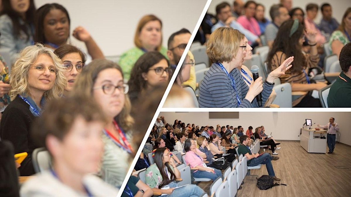
The Spectral Flow Cytometry Symposium has a strong focus in ensuring delegates have a highly interactive experience :
- Listen to talks from international speakers detailing the spectral cytometry technology and analysis methods
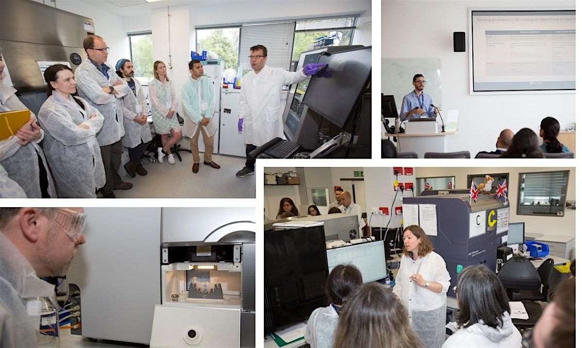
- Participate in hands-on workshops to explore a range of cutting-edge equipment and analysis techniques
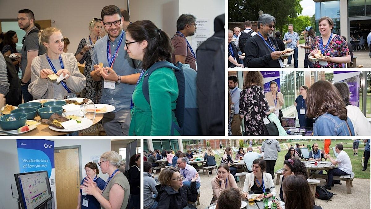
- Meet with technical specialists from leading flow cytometry companies, explore a range of cutting-edge equipment offered by exhibiting companies.
- Explore an extensive exhibition featuring cutting-edge equipment from leading companies
- Enjoy great food throughout the symposium and connect with the community – plus an evening social on 1st July
Invited speakers:
Sarah Bonte - Postdoc, VIB-UGent, Belgium
"Workflows for comparing spectral flow cytometry data across multiple technologies"
Jessica Back - Director, MICR Core & Research Core Facilities, Wayne State University, USA
"A Robust Pairing: Spectral Cytometry and High Parameter Imaging"
Ruth Barnard - Director, GSK, UK
"Necessity is the Mother of Innovation: Spectral Flow Cytometry in a GCP Laboratory"
Thomas Hayday - Chief Scientific Officer & Founder, IMU Biosciences, UK
"Mapping the Future of Medicine: Using Spectral Cytometry and AI to Decode the Human Immune System"
Florian Mair - Director Flow Cytometry Facility, ETH Zurich, Switzerland
"Elucidating the complexity of spread in the era of 50-color spectral cytometry"
Bartek Rajwa - Purdue University, USA
"Making sense of panel-quality metrics in spectral cytometry: a practical statistical view"
Andy Riddell - Head of Flow Cytometry, Francis Crick Institute, UK
"Cytometry: What Has Changed, What Hasn’t, and What It Means for Expertise"
Programme:
Wednesday 1st July Registration with tea & coffee is open from 8.30am
9:15 – 9.40: Welcome from Rachael Walker, Head of Flow Cytometry, Babraham Institute
9:40 – 10:20: A Robust Pairing: Spectral Cytometry and High Parameter Imaging - Jessica Back, Director, MICR Core & Research Core Facilities, Wayne State University, USA
10:20 – 11.00: Necessity is the Mother of Innovation: Spectral Flow Cytometry in a GCP Laboratory - Ruth Barnard, Director, GSK, UK
11.00- 11:30 Refreshments & Exhibition kindly supported by Dotmatics
11:30 – 13.00 WORKSHOP SESSION 1
13.00 – 14.00 Lunch kindly supported by Slingshot Biosciences
14.00 – 15:30 WORKSHOP SESSION 2
15:30 – 16.00 Refreshments & Exhibition
16.00 – 16:40: Cytometry: What Has Changed, What Hasn’t, and What It Means for Expertise - Andy Riddell, Head of Flow Cytometry, The Francis Crick Institute, UK
16:40 - 17:20: Elucidating the complexity of spread in the era of 50-color spectral cytometry - Florian Mair, Director Flow Cytometry Facility, ETH Zurich, Switzerland
17.20-17.40 Closing – polls
18:00 Evening Social kindly supported by Sony
Thursday 2nd July
9:00 - 9:05 Welcome back to Day Two, Rachael Walker, Head of Flow Cytometry, Babraham Institute
9:05 – 9:45: Workflows for comparing spectral flow cytometry data across multiple technologies - Sarah Bonte, Postdoc, VIB-UGent, Belgium
9:45 – 10:25: Mapping the Future of Medicine: Using Spectral Cytometry and AI to Decode the Human Immune System - Thomas Hayday, Chief Scientific Officer & Founder, IMU Biosciences
10:25 - 11:00 Refreshments & Exhibition
11:00 – 12:30 WORKSHOP SESSION 3
12:30-13:30 Lunch kindly supported by FluoroFinder
13:30 – 15:00 WORKSHOP SESSION 4
15:00 - 15:30 Refreshments & Exhibition
15:30 – 16:10: Making sense of panel-quality metrics in spectral cytometry: a practical statistical view - Bartek Rajwa, Research Professor, Purdue University, USA
16.10- 16.30 Closing Address Polls
🌈 Before You Unmix: A Beginner’s Introduction to Spectral Flow course
Course tutors: Rachael Walker and Derek Davies
30th June 2026 – 13:00-17:00
If you’re new to spectral flow cytometry, this half‑day course is the perfect place to begin. We’ll break down the essentials in a simple, friendly way — no jargon, no assumptions, no overwhelm. Together, we’ll explore what “full spectrum” really means, why spectral flow works differently from conventional flow, and how unmixing transforms raw data into something you can actually interpret.
Whether you’re taking this session as a standalone introduction or using it to get comfortable before diving into the symposium, you’ll leave with a clear understanding of the core concepts, the confidence to follow more advanced discussions, and the sense that spectral flow isn’t nearly as mysterious as it first appears. Think of it as the smooth on‑ramp to the spectral highway — helping you feel prepared, empowered, and ready to get the most out of everything that follows.
· Gain a clear understanding of the principles behind full‑spectrum detection.
· Learn how fluorochrome signatures contribute to unmixing.
· Identify the key reference controls required for reliable spectral experiments.
· Interpret simple spectral plots and unmixing outcomes.
· Enter the symposium with a solid, foundational understanding of the terminology and workflows.
Symposium Workshops
Thanks to the unique set up at the Babraham Institute, the Symposium is able to offer interactive workshops on a range of commercially available spectral cytometers.
A brand new selection of workshop sessions will be offered to delegates in 2026. Each delegate will be able to choose 4 workshops to attend over the two days. Delegates will be emailed 2 weeks before the Symposium and asked to select their workshop choices.
This year the Symposium's workshops will include a range of practical and classroom based topics.
2026 Workshops
Workshop Titles and abstracts
Lab based: We are able to offer a practical sessions on each of the following instruments – NB not all instruments are available for each time slot, so please look at the time table.
- BD FACSDiscover S8 Spectral and Imaging Sorter
- Cytek Aurora CS Spectral Sorter
- Thermo Fisher Bigfoot Spectral Sorter
- Sony ID7000 Spectral Analyser
- Cytek Aurora Evo Spectral Analyser
- Thermo Fisher Attune Xenith Spectral Analyser
- Beckman Coulter CytoFLEX mosaic Spectral Analyser
- Agilent NovoCyte Opteon Spectral Analyser
- BD FACSDiscover A8 analyser
- ChallenBio Cytostellar Spectral Analyser
- The Slingshot Effect: Using synthetic cells to gain real precision.
Classroom-based
Preparing Spectral Flow Cytometry Data for Publication: Practical Statistical Analysis and Presentation
Bartek Rajwa
This introductory, interactive workshop will cover fundamental principles for analyzing, presenting, and publishing spectral flow cytometry data. Although designed to be accessible to participants with limited statistical, mathematical, or programming background, the workshop will also be useful for advanced users because it will explain, in depth, the basic quantitative operations underlying spectral cytometry. These will include the mathematical logic of spectral unmixing, the computation and interpretation of spectral panel-quality metrics, and the first-principles reasoning behind operations that are often treated as black boxes.
The goal is to make these operations transparent: what is being calculated, why it is calculated, what assumptions are involved, and how the resulting values should be interpreted in the context of publishable cytometry data. All key concepts will be demonstrated using simple R code and hands-on examples, allowing participants to follow each calculation step by step.
The workshop will also address practical issues in statistical presentation, including how to display compositional data such as cell-population percentages, why transformations are often used for visualization, and how to distinguish effect size from statistical significance. Particular emphasis will be placed on why p-values cannot be used to communicate the magnitude or biological relevance of an observed effect. Participants are encouraged to bring their own laptop computers with R installed so they can follow along with the demonstrations and exercises.
Staining success: blocking, titration and fixation
Oliver Burton, University of Cambridge
Sample preparation is key to reproducible, high-quality flow cytometry data. In this workshop, I'll cover a range of tips and tricks to improve signal and reduce background in flow cytometry assays. Topics will include antibody titration, different types and purposes of blocking reagents, types of fixatives and when to use them, and a discussion on how these factors influence panel design and stability.
Best practices in high-dimensional spectral panel design: a step-by-step guide to success
Florian Mair, Eth Zurich
Recent advances in cytometry instrumentation and reagents have increased the maximum possible dimensionality of a fluorescence-based flow cytometry experiment up to 50 different markers. However, without appropriate guidance, designing these high-dimensional panels can be time-consuming and frustrating.
In this workshop, we outline systematic workflows to design any panel from 10-50 parameters on conventional and spectral cytometers, while minimizing wet-lab trial and error. After covering theoretical aspects of spectral flow cytometry and practical basics that are relevant for panel design, we will use an interactive format to design a panel and discuss typical caveats and problems. Specifically, we will: Summarise key aspects of how spectral flow cytometry works
Computational Cytometry 101
Sarah Bonte, VIB-UGent Center for Inflammation Research, Belgium
The default computational analysis pipeline consists of three main steps: pre-processing and quality control, clustering, and downstream (statistical) analysis. Quality control (i.e. removing low quality events and detecting and solving batch effects) is especially important in a computational setting, because of the garbage in, garbage out principle. The overall pipeline (and specific considerations for spectral data) will be discussed and some computational tools like PeacoQC for quality control, CytoNorm for batch effect removal and FlowSOM for clustering will be highlighted.
Approaches to Autofluorescence (AF) Management with Full Spectrum Profiling™ (FSP) and Tools within SpectroFlo® to Enhance Resolution in Samples with Complex AF.
Adam Davison, Cytek Biosciences
Characterizing and understanding the impact of autofluorescence (AF) in our samples allows for the determination of suitable methodologies to mitigate its impact upon antigen resolution. This workshop will introduce how to qualify AF impact and top select suitable approaches including panel design recommendations to optimize end outcomes. Here we will showcase existing tools within SpectroFlo® to rapidly overcome intense and complex AF in minutes using an example case study.
Advanced Analysis in OMIQ: Secure Collaboration and Parallel Algorithms in the Cloud
Jamie McCarthy, FCS Express, Dotmatics
OMIQ (www.omiq.ai) offers an innovative solution bridging both classical and high-dimensional analysis in a pleasantly designed cloud software which also allows collaboration, automation, and data management. You can expect to see an overview of the OMIQ platform, including our integrated features. We will also run a live demonstration of Data Analysis in OMIQ; going from raw data to p-values. Through our integration with Graphpad Prism you can also reduce manual data handling and automate your statistical analysis.
Spectral Cytometry is enabling more parameters, more files and more events in your experiments and our OMIQ Application Scientist will focus on modern high parameter algorithms and solutions hosted in a cloud environment to get the most out of your data.
Approaches to Spectral Unmixing Challenges: A Detective's Journey
Laura Ferrer Font, Waters Biosciences
The overarching goal of this workshop is to provide all participants with the knowledge and tools they need to successfully design and troubleshoot their polychromatic cytometry experiments irrespective of the platform used.
Supporting Spectral Cytometry: An SRL Perspective
Jessica Back, Wayne State University, USA & Philip Hobson, Francis Crick Institute, London
This workshop will focus on real-world experiences with integrating spectral technologies into a Shared Resource Laboratory (SRL). Discussion topics will include:
Developing staff expertise
Managing complex analysis pipelines
Data management and storage
Managing user expectations
Ask the Experts Q&A session
Rachael Walker, Oliver Burton, Jessica Back, Phil Hobson
Moderator: Derek Davies
Although Spectral Flow Cytometry is now an established technology and has been adopted by many Core Labs and researchers, there still remain challenges. This moderated session with bring together experts to address questions that delegates may have related to hardware, experimental design best practices, data analysis, reagents, and more.
From Unmixing to High-Dimensional Insights in FlowJo v10
Serena di Cecilia
Spectral data can feel complex but with the right workflow, it becomes a powerful discovery tool. This workshop takes participants from spectral unmixing and quality control through to high-dimensional data analysis in FlowJo v10. By exploring tools such as UMAP, t-SNE, and clustering, attendees will learn how to uncover hidden populations and turn complex datasets into clear, interpretable results.
From Unmixing to High-Dimensional Insights in FlowJo v11
Serena Di Cecilia
Spectral data can feel complex but with the right workflow, it becomes a powerful discovery tool. This workshop takes participants from spectral unmixing and quality control through to high-dimensional data analysis in FlowJo v11. By exploring tools such as UMAP, t-SNE, and clustering, attendees will learn how to uncover hidden populations and turn complex datasets into clear, interpretable results.
Empower spectral data analysis with the Cytobank platform: Automated gating, Advanced illustration and Unsupervised Discovery
Maria Salvany
Spectral flow cytometry produces highly complex datasets that require powerful analytical tools to unlock their full biological value. This workshop introduces Cytobank's end-to-end platform for spectral data analysis, featuring automated gating, integrated statistics, flexible publication-ready visualizations, and advanced machine learning algorithms for unsupervised population discovery. Attendees will learn practical approaches for dimensionality reduction, clustering, biomarker discovery, and reproducible analysis workflows that accelerate insight generation from high-dimensional cytometry experiments.
Further Symposium Information
Tickets will provide delegates with full access to the Sympsoium for both days and a choice of four workshops (bookings for the focused Symposium's workshops will be offered to registered delegates at a later date).
Discounted tickets are available for academic staff, clinical centres, Facility Cores/Shared Resource Laboratories and Babraham Research Campus attendees.
This event is kindly sponsored by
Where is it happening?
Event Location & Nearby Stays:
GBP 102.00