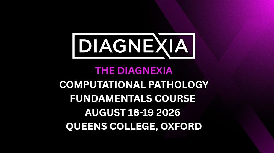
The Diagnexia Computational Pathology Course 2026
About this Event
Diagnexia Computational Pathology Course
📍 Queens College, Oxford
📅 Date: August 18 and 19 2026
August 18th Accomodation Included With Ticket Purchase
Get full access to both the Symposium and the Computational Pathology Course at a discounted rate.
Bundle price includes:
- Consultant discounted rate: £475 (previous rate for both £560)
- Trainee rate: £327 (previous rate for both £385)
Limited availability.
Day 1
09:30 – 10:00 Registration & Coffee
Sign-in, materials collection, networking
10:00 – 10:30 - Welcome & Introduction to Computational Pathology - RC + PM
Course overview and learning objectives; definition and scope of computational pathology; distinction from digital pathology; the data-intensive nature of pathology; course roadmap
10:30 – 11:15 - Digital Image Fundamentals I: Core Concepts - PM
What is a digital image; pixels, resolution, and bit depth; color spaces (RGB, HSV, LAB); relationship between magnification and microns per pixel (mpp); image file formats (JPEG, PNG, TIFF); lossy vs. lossless compression and their impact on analysis; why format choices matter downstream
11:15 – 11:30 - Coffee Break
11:30 – 12:15 - Digital Image Fundamentals II: Whole-Slide Imaging - PM
Whole-slide imaging technology; scanner types and acquisition; pyramidal/multi-resolution image structure; proprietary vs. open WSI formats (SVS, NDPI, DICOM WSI); storage, memory, and bandwidth considerations; practical implications for computational pipelines
12:15 – 13:00 - Stain Physics and Color Analysis - AJ
Physics of stain absorption (Beer-Lambert law); H&E and special stains from a computational perspective; stain vectors and color deconvolution; stain normalization approaches; quantification of chromogenic and fluorescent signals; inter-laboratory variability challenges
Lunch - 13:00 – 14:00
14:00 – 14:45 - Image Processing Fundamentals - AJ
Intensity transformations; histogram processing; spatial filtering (smoothing and sharpening); morphological operations (erosion, dilation, opening, closing); practical applications in tissue analysis
14:45 – 15:30 - Segmentation in Pathology - PM
Tissue vs. background detection; thresholding methods (global, Otsu’s, adaptive); region-based and edge-based approaches; nuclear and cell segmentation; the reference area problem
15:30 – 15:45 - Coffee Break
15:45 – 16:30 - Feature Extraction: Classical Approaches - AJ
Morphological features (size, shape, texture); nuclear and cellular descriptors; glandular and architectural features; tumor microenvironment metrics; building interpretable feature sets
16:30 – 17:15 - Introduction to Machine Learning for Pathology - AJ
Supervised vs. unsupervised learning; training, validation, and test sets; classical methods (random forests, SVM); performance metrics and their clinical interpretation; avoiding common pitfalls
17:15 – 18:00 - Introduction to Deep Learning - AJ
Neural network fundamentals; forward propagation and backpropagation (conceptual); activation functions; loss functions and optimization; regularisation techniques; training vs. inference; why deep learning transformed computational pathology
18:00 – 18:15 - Day 1 Wrap-up and Q&A
Summary of key concepts; preview of Day 2; open questions
Day 2
08:30 – 09:00 Coffee & Networking
09:00 – 09:15 - Day 1 Recap and Day 2 Overview - PM
Brief review of foundational concepts; outline of advanced topics
09:15 – 10:00 - Deep Learning Architectures for Pathology - EK
Convolutional neural networks (CNNs) explained; landmark architectures (ResNet, U-Net); Vision Transformers (ViT); classification, segmentation, and detection networks; transfer learning and domain adaptation
10:00 – 10:45 - Training Strategies and Data Challenges - AJ
Data preparation and patch extraction; annotation strategies and quality; handling class imbalance; data augmentation (geometric, color, stain); managing staining and scanner variability
10:45 – 11:00 - Coffee Break
11:00 – 11:45 - Multiple Instance Learning and Slide-Level Prediction - JW
The MIL paradigm for whole-slide analysis; attention-based aggregation; weakly supervised learning; from patches to patient-level predictions
11:45 – 12:30 - Foundation Models and Vision-Language Models - EK
Self-supervised and contrastive learning; pathology foundation models (overview); vision-language models (CLIP, PathChat, CONCH); zero-shot classification; conversational AI for pathology; limitations and hallucination risks
12:30 – 12:45 - Morning Q&A
Lunch - 12:45 – 13:45
13:45 – 14:30 - Clinical Applications I: Diagnostic Support - AJ
Cancer detection and classification (breast, prostate, colorectal, lung); Gleason grading automation; metastasis detection; quality control and triage applications; discussion on clinical utility
14:30 – 15:15 - Clinical Applications II: Biomarkers and Prognosis - JW
IHC quantification (Ki-67, HER2, PD-L1); tumor microenvironment analysis; TIL assessment; predicting molecular features from H&E survival and treatment response prediction; discussion on clinical utility
15:15 – 15:30 - Coffee Break
15:30 – 16:00 - Validation, Regulation, and Deployment - JA
Evaluation metrics for classification, segmentation, detection; cross-validation and external validation; regulatory pathways (FDA, CE/MDR); algorithmic fairness and bias considerations; clinical integration and continuous monitoring
16:00 – 16:30 - Model Interpretability and Explainable AI - EK
Why interpretability matters in clinical pathology; XAI techniques (saliency maps, Grad-CAM, attention visualization); accuracy vs. interpretability trade-offs; building pathologist trust
16:30 – 17:00 - Emerging Technologies - AJ
Multiplex imaging analysis (mIF, IMC, spatial transcriptomics); multimodal data integration; federated learning; AI-augmented pathology workflows
17:00 – 17:30 - Future Directions and the Pathologist’s Role - PM
The pathologist’s role in computational pathology development; integration of imaging with -omics data; AI-assisted reporting and case management; building multidisciplinary collaborations; career pathways and skill development
17:30 – 18:00 - Open Discussion, Q&A, and Closing
Panel discussion; participant questions; key takeaways; resources for continued learning; course evaluation; closing remarks
Speakers abbreviations
RC - Prof. Runjan Chetty Chief Medical Officer at Deciphex/Diagnexia
EK - Emre Köse Computational Pathologist @ Deciphex
AJ - Andrew Janowczyk Assistant Professor at Emory University
JW - John Weldon Clinical AI Director @ Deciphex
JA - Jonathan Armstrong AI Governance Lead @ Deciphex
PM - Pierre Moulin MD, PhD — Chief Scientific Officer @ Deciphex
Where is it happening?
Event Location & Nearby Stays:
GBP 119.89 to GBP 515.32